The Hidden Side of Exocad: 6 Ways to Maximize Its Power
Exocad is undoubtedly one of the most powerful design software tools out there.
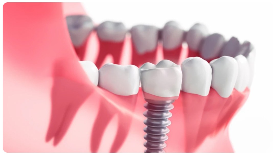
Surgical Guide Implant: A Seamless and Precise Approach to Implant Dentistry
Dentistry is continuously evolving with technological advancements, offering more comfortable, safe, and successful treatment methods for patients.(Akova & Üstün, 2006) One of the innovations that has gained popularity in recent years is the seamless implant treatment. Surgical Guide Implant (Guided Flapless and Stitch-Free Implant Technique) is a modern approach supported by CAD/CAM technologies, minimizing errors through patient-specific planning.(İkiz et al., 2023)
In traditional implant methods, the gum is incised, the implant is placed, and then stitches are applied, followed by a healing process. However, in the stitch-free implant technique, the treatment location, application angle, and depth are determined using intraoral and bone structure imaging without surgical incisions. The main advantages of this method are:
Seamless implant surgery requires detailed analysis and planning. The steps involved are as follows:
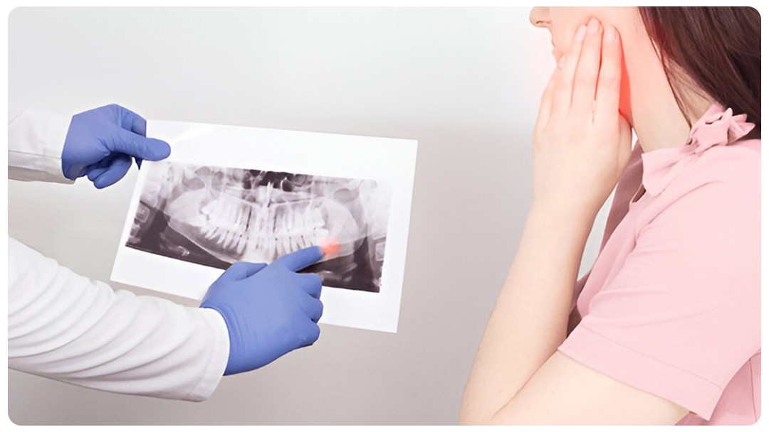
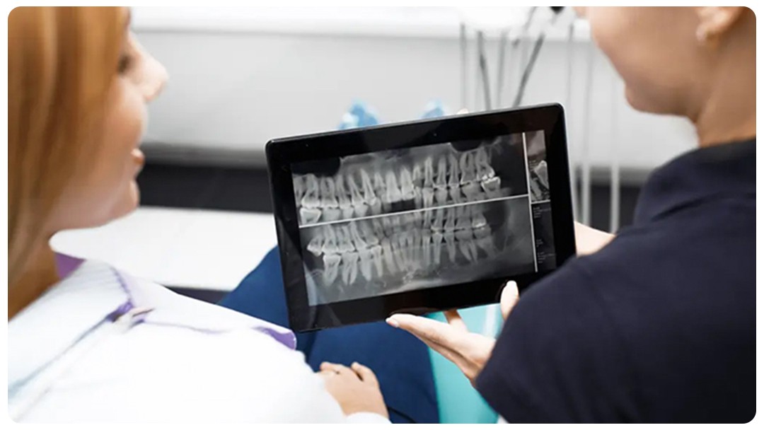
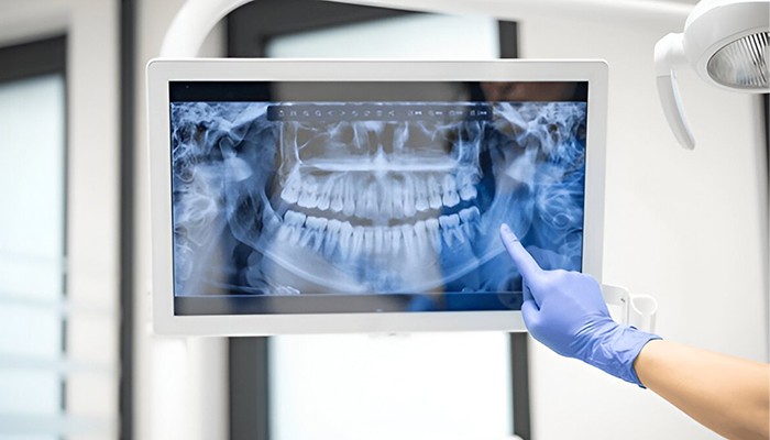
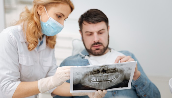
Standard panoramic X-rays provide two-dimensional images; however, for the stitch-free implant technique, a detailed evaluation of the jawbone is essential.(Akova & Üstün, 2006) Therefore, Cone Beam Computed Tomography (CBCT) is used for three-dimensional imaging. This process enables:

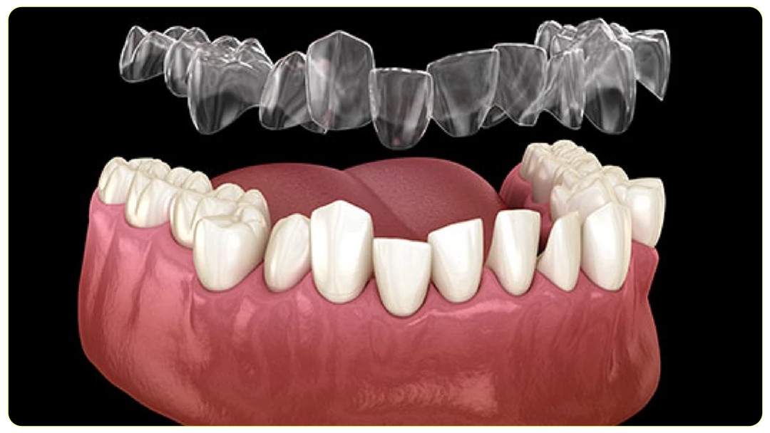
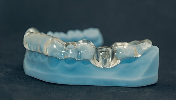
In the stitch-free implant technique, a custom-made surgical guide is used. The Implant Guide is a template that pre-determines where and at what angle the implant should be placed. With this guide:
Also referred to as a closed surgical technique, this method involves implant placement without incising the gum. Thanks to the surgical guide, the procedure ensures:(Akova & Üstün, 2006)

This method is applicable to many patients with a suitable jawbone structure. Due to its advantages, it is particularly preferred in the following cases:(İkiz et al., 2023)

| Feature | Traditional Implant | Stitch-Free Surgical Guide Implant |
| Surgical Incision | Required | Not Required |
| Healing Time | Longer | Faster |
| Pain and Swelling | Can be significant | Minimal |
| Treatment Duration | Longer | Shorter |
| Precision | Dependent on surgeon’s expertise | High precision with 3D planning |
In conclusion
Surgical Guide stitch-free implant treatment is a comfortable option for patients and a safe, precise method for dentists. The advanced 3D imaging technology and surgical guides make this process significantly more successful.