The November 19, 2026 release of Grand Theft Auto VI is one of the most talked-about topics right now. The generation counting down to launches like this grew up with screens, instant feedback and visual explanations. It is hardly surprising that the same people, once they sit in the dental chair, want to see what is happening, understand it and skip unnecessary waiting.
In the clinic, that expectation usually meets reality first at the impression stage. An intraoral scanner, a 3D model that builds up on the screen, and a provisional crown, splint or working model printed from that file are changing the patient experience step by step. In this article we look at how patients experience scanning, what the on-screen model adds to communication, how a scan file becomes a 3D print, and where the digital workflow still reaches its limits, based on peer-reviewed publications with DOIs.
Why do patients find intraoral scanning more comfortable than conventional impressions?
An intraoral scanner is a device that uses light to capture a three-dimensional digital model of the teeth and surrounding tissues directly in the mouth. With a conventional impression, the patient waits while impression material in a tray sets in the mouth. According to the review by Mangano et al. (2017), some patients, such as those with a strong gag reflex and children, tolerate the conventional procedure poorly, and removing the tray and impression material is a clear advantage for them.
Patient-reported findings generally point in the same direction:
Siqueira et al. (2021) reviewed 17 clinical studies in fixed prosthodontics and implant dentistry, covering 437 patients. Scanning was generally faster than conventional impressions regardless of the size of the scanned area or whether the restoration was tooth- or implant-supported, and patients generally preferred it. Prosthodontic outcomes were similar with both workflows.
Joda and Brägger (2016) compared both methods in the same 20 patients receiving single implant crowns. On a 0-100 scale, the convenience score was 78.6 for scanning and 53.6 for conventional impressions. All patients said they would choose the digital workflow if given the choice in the future.
Christopoulou et al. (2022) reviewed nine studies in orthodontic patients. Experiences with intraoral scanners were generally more positive regarding smell, taste, nausea and gagging. However, anxiety levels did not differ between the methods, and how patients perceived the duration varied between studies.
Garcovich et al. (2026) pooled seven studies with 337 children. Scanning was faster, and children reported less pain, less breathing difficulty, less gag reflex and greater comfort. Children were more than four times as likely to prefer scanning.
In short, for most patients scanning is a more comfortable experience than waiting for a material to set in their mouth. It is still worth remembering that anxiety does not disappear and that not every patient perceives the procedure as short.
What changes when patients see a 3D model of their own teeth on screen?
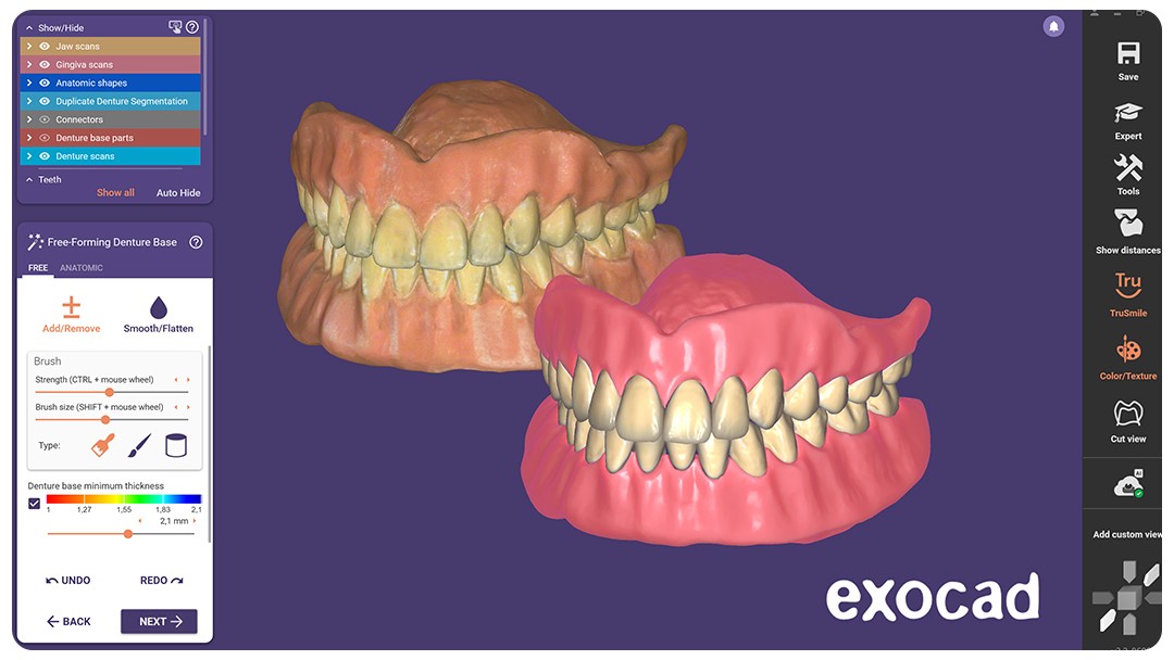
With a conventional impression, the patient often does not see a result until the stone model comes back from the lab. With scanning, the model builds up on the screen during the procedure, and the dentist can explain findings using the same image. Mangano et al. (2017) note that patients feel more involved in their treatment and that communication with them becomes more effective. According to the authors, this sense of involvement may have a positive effect on the treatment, for example on oral hygiene compliance.
The first randomized clinical trial on this question comes from pediatric dentistry. Schulz-Weidner et al. (2024) explained the dental findings and treatment plans of 60 children to their parents in one of two ways: verbally only, or verbally with the intraoral scan as a visual aid. In the questionnaire that followed, 85.5% of answers on average were correct in the group shown the scan, compared with 57.2% in the verbal-only group. The largest differences were in questions about treatment needs and planned therapy. The authors describe it as a first pilot study.
Baradee and Spies (2025) point to another benefit of digital records: scans can be archived and compared directly with later scans, which helps monitor changes such as wear or tooth movement over time. For the patient, this means the question "what has changed since my last visit?" has an answer they can actually see.
How does a scan file become a 3D-printed provisional crown, splint or model?
What you get at the end of a scan is a file. Mangano et al. (2017) describe how the digital model can be sent to the laboratory in a proprietary format or as an STL file, removing the need to pour stone casts or ship impressions by courier. The steps that follow look broadly like this:
Scan and check: The dentist or technician reviews the model on screen. Mangano et al. (2017) highlight that the technician can check the file right away and request a new scan while the patient is still in the chair, which can save a second appointment.
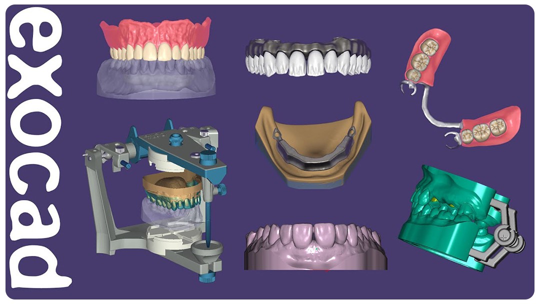
Design: The file is imported into CAD software, where the crown, splint or model is designed. Tian et al. (2021) describe 3D printing as producing personalized objects from digital models created with computer-aided design.
Print preparation: The design is loaded into the printer software, and the build angle and supports are set. For details, see our articles on build orientation in dental 3D printing and support structures.
Printing: The part is printed layer by layer from a light-cured resin. Revilla-León and Özcan (2019) review the main additive manufacturing processes used for polymers in prosthetic dentistry and their applications. The right settings depend on the printer and resin; the manufacturer's instructions come first.
Post-processing: The printed part is washed, dried and post-cured. We covered these steps in our guides on washing dental 3D prints and overcure and undercure (in Turkish).
Try-in and delivery: The part is tried in the mouth or on the model and adjusted if needed.
The same file can lead to different devices. For fit and strength of printed provisionals, see 3D-printed provisional crowns and bridges; for night guards, 3D-printed occlusal splints; and for aligner models, 3D-printed models for clear aligners.
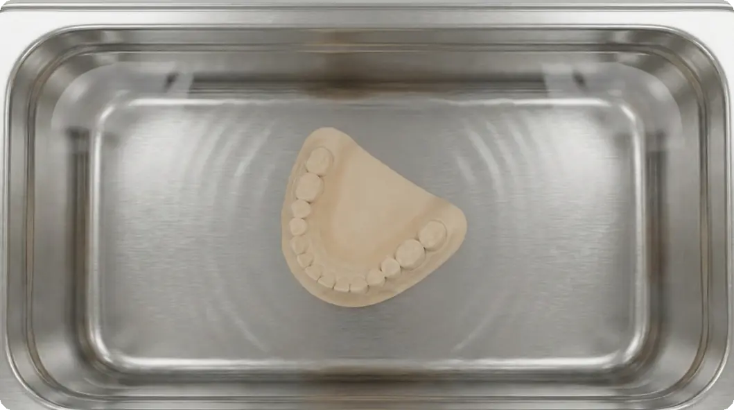
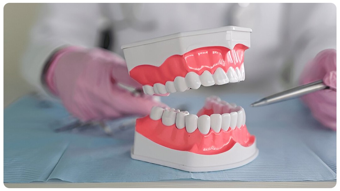
Figure 1: 3D-printed dental model
Is faster always better? What are the limits of the digital workflow?
The digital workflow saves time in many cases, but not in every case. The limits most often reported in the literature are:
Subgingival margins: A scanner can only record what it can see. Mangano et al. (2017) note that deep margin lines on prepared teeth and bleeding are difficult to capture with intraoral scanners. In a laboratory study by An et al. (2024), finish lines placed 0.5 and 1.0 mm subgingivally were scanned with greater discrepancies than equigingival finish lines. Saliva contamination also significantly increased discrepancies, and the effect was larger for subgingival margins.
Long spans and movable tissues: Mangano et al. (2017) state that the literature up to that point did not support the use of intraoral scanners for long-span restorations. Kihara et al. (2020) describe cross-arch fixed prostheses as difficult to fabricate from intraoral scans, and Baradee and Spies (2025) list movable oral tissues, deep subgingival margins and long-span prostheses as situations where digital impressions still face challenges.
Time for complete-arch scans: In a randomized clinical trial by Sailer et al. (2019) in 10 patients needing a bridge, complete-arch scanning took longer than the conventional impression with all three scanners tested: 658 seconds on average for the conventional impression versus 1091 to 1702 seconds for the scans. Some of the scanners in that trial required powdering the teeth, and patients did not clearly prefer any method. On the other hand, the review by Siqueira et al. (2021), covering 17 clinical studies, found scanning generally faster than conventional impressions, including complete-arch scans. Results can vary with the device and the case.
Learning curve: Mangano et al. (2017) point out that introducing a scanner into the clinic involves a learning curve.
Steps after printing: A printed part is not ready to use the moment it leaves the printer. Tian et al. (2021) note that post-processing takes time. Washing and post-curing should never be skipped.
So the speed digital-native patients expect is real, but it has limits. Careful case selection, a clean and dry working field and complete post-processing are what make speed and quality go together.
Frequently asked questions
Does intraoral scanning hurt?
Scanning uses light rather than impression material. In the meta-analysis by Garcovich et al. (2026), children reported less pain and greater comfort with scanning than with conventional impressions.
Is scanning better for people with a strong gag reflex?
Mangano et al. (2017) note that patients with a strong gag reflex can find conventional impressions hard to tolerate, and that removing the tray and material is an advantage for them. Christopoulou et al. (2022) also found that nausea and gagging were generally reported less often with scanners.
Can an intraoral scanner replace conventional impressions in every case?
No. Deep subgingival margins, bleeding, saliva, movable tissues and long-span bridges are among the situations where scanning still struggles (Mangano et al., 2017; An et al., 2024; Baradee and Spies, 2025). The dentist chooses the method for each case.
How does a scan file get to the 3D printer?
The scan is exported as a file such as STL, designed as a crown, splint or model in CAD software, then oriented, supported and printed in the printer software. The printed part is used after washing and post-curing.
Does seeing a model of my teeth on screen actually help?
In the study by Schulz-Weidner et al. (2024), parents who were shown the scan while findings were explained answered questions about treatment needs and plans correctly much more often than parents who received a verbal explanation only.
Are my digital records useful later on?
Yes. Baradee and Spies (2025) note that digital records can be archived and compared with later scans to monitor treatment over time.
References
Mangano F, Gandolfi A, Luongo G, Logozzo S. Intraoral scanners in dentistry: a review of the current literature. BMC Oral Health. 2017;17(1):149. https://doi.org/10.1186/s12903-017-0442-x
Siqueira R, Galli M, Chen Z, Mendonça G, Meirelles L, Wang HL, et al. Intraoral scanning reduces procedure time and improves patient comfort in fixed prosthodontics and implant dentistry: a systematic review. Clinical Oral Investigations. 2021;25(12):6517–6531. https://doi.org/10.1007/s00784-021-04157-3
Joda T, Brägger U. Patient-centered outcomes comparing digital and conventional implant impression procedures: a randomized crossover trial. Clinical Oral Implants Research. 2016;27(12):e185–e189. https://doi.org/10.1111/clr.12600
Christopoulou I, Kaklamanos EG, Makrygiannakis MA, Bitsanis I, Tsolakis AI. Patient-reported experiences and preferences with intraoral scanners: a systematic review. European Journal of Orthodontics. 2022;44(1):56–65. https://doi.org/10.1093/ejo/cjab027
Garcovich D, Aiuto R, Lorenzo AA, Bouserhal J, Dioguardi M, Martin MA. Digital Versus Conventional Impression Techniques in Children: A Systematic Review and Meta-Analysis of Efficiency, Comfort and Patient Preference. International Journal of Paediatric Dentistry. 2026;36(2):254–264. https://doi.org/10.1111/ipd.70045
Schulz-Weidner N, Gruber M, Schraml EM, Wöstmann B, Krämer N, Schlenz MA. Improving the Communication of Dental Findings in Pediatric Dentistry by Using Intraoral Scans as a Visual Aid: A Randomized Clinical Trial. Dentistry Journal. 2024;12(1):15. https://doi.org/10.3390/dj12010015
Baradee AA, Spies BC. Are Conventional Impressions Obsolete? A Narrative Review on the Applicability of Intraoral Scanners. Australian Dental Journal. 2025;70(Suppl 1):S93–S104. https://doi.org/10.1111/adj.70015
Tian Y, Chen C, Xu X, Wang J, Hou X, Li K, et al. A Review of 3D Printing in Dentistry: Technologies, Affecting Factors, and Applications. Scanning. 2021;2021:9950131. https://doi.org/10.1155/2021/9950131
Revilla-León M, Özcan M. Additive Manufacturing Technologies Used for Processing Polymers: Current Status and Potential Application in Prosthetic Dentistry. Journal of Prosthodontics. 2019;28(2):146–158. https://doi.org/10.1111/jopr.12801
An H, Mickesh GJ, Cho D, Sorensen JA. Effect of finish line location and saliva contamination on the accuracy of crown finish line scanning. Journal of Prosthodontics. 2024;33(1):86–94. https://doi.org/10.1111/jopr.13658
Kihara H, Hatakeyama W, Komine F, Takafuji K, Takahashi T, Yokota J, et al. Accuracy and practicality of intraoral scanner in dentistry: A literature review. Journal of Prosthodontic Research. 2020;64(2):109–113. https://doi.org/10.1016/j.jpor.2019.07.010
Sailer I, Mühlemann S, Fehmer V, Hämmerle CHF, Benic GI. Randomized controlled clinical trial of digital and conventional workflows for the fabrication of zirconia-ceramic fixed partial dentures. Part I: Time efficiency of complete-arch digital scans versus conventional impressions. The Journal of Prosthetic Dentistry. 2019;121(1):69–75. https://doi.org/10.1016/j.prosdent.2018.04.021